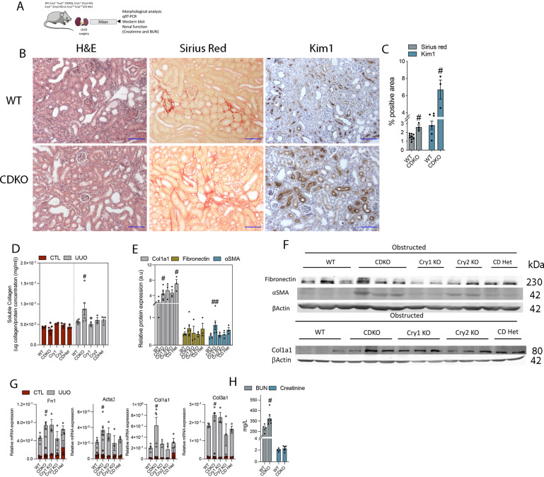
Figure 3. Fibrosis is exacerbated by Cry 1/2 genetic deficiency.
(A) Schematic of experimental design of WT and Cry1/Cry2 KO mice subjected to unilateral ureteral obstruction (UUO) for 3 d. (B) Representative microphotographs of H&E, Sirius red, and KIM1 immunohistochemistry stains from obstructed kidneys of WT and CDKO. Scale bar: 150 μm. (C) Quantification of Sirius red and KIM1 immunohistochemistry in kidney sections of WT and CDKO mice (Sirius red number of mice: WT [8], CDKO [3]; KIM1 number of mice: WT [6], CDKO [3]). (D) Bar plot represents the quantification of the total soluble collagen in frozen kidneys from WT, CDKO, Cry1 KO, Cry2 KO, and CDHet mice. (E, F) Bar plot represents the relative protein expression of Col1a1, fibronectin, and SMA of images in (F). (F) Immunoblot depicting the expression of fibronectin, αSMA, and Col1a1 in kidneys from WT, CDKO, Cry1 KO, Cry2 KO, and CDHet mice subjected to UUO for 3 d, β-actin levels were used for normalization. (G) Relative mRNA expression of fibrosis-related genes in WT, CDKO, Cry1 KO, Cry2 KO, and CDHet mice subjected to UUO. (H) BUN and plasma creatinine levels of WT and CDKO mice 3 d after UUO. Number of mice: Cry-deficient mice: WT (n = 6), CDKO (n = 6), Cry1 KO (n = 3), Cry2 KO (n = 3), CDHet mice (n = 3); #P < 0.05, ##P < 0.01 compared with the WT (Mann–Whitney).
Source data are available for this figure.

