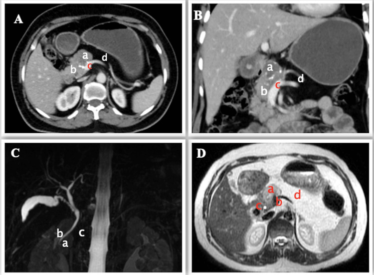
Figure 1. Preoperative imaging.
A, B - CECT (axial and coronal view) scan showing SPEN (a) arising from the head of pancreas (b) with infiltration of the portal vein (c) and agenesis of dorsal pancreas (d)
C - MRCP showing normal ventral pancreatic duct (of Wirsung) (a), the common bile duct (b), and absence of dorsal pancreatic duct (c)
D - MRI showing tumor (a) just above the portal confluence (b), close by distal CBD (c) and distal pancreas replaced by fat (d)
CECT: contrast-enhanced computed tomography, SPEN: solid pseudopapillary epithelial neoplasm, MRCP: magnetic resonance cholangio-pancreatography, CBD: common bile duct

