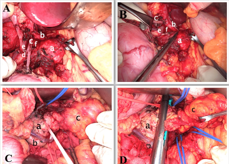
Figure 2. Intraoperative photos.
A, B - Tumor (a) freed from common hepatic artery (b) and gastroduodenal artery (c), posterior superior pancreaticoduodenal artery (clipped) (d), CBD (e), portal vein (f)
C, D - Showing looping and transection of the head of the pancreas (a), duodenum (b), dorsal pancreas replaced by fat (c)
CBD: common bile duct

