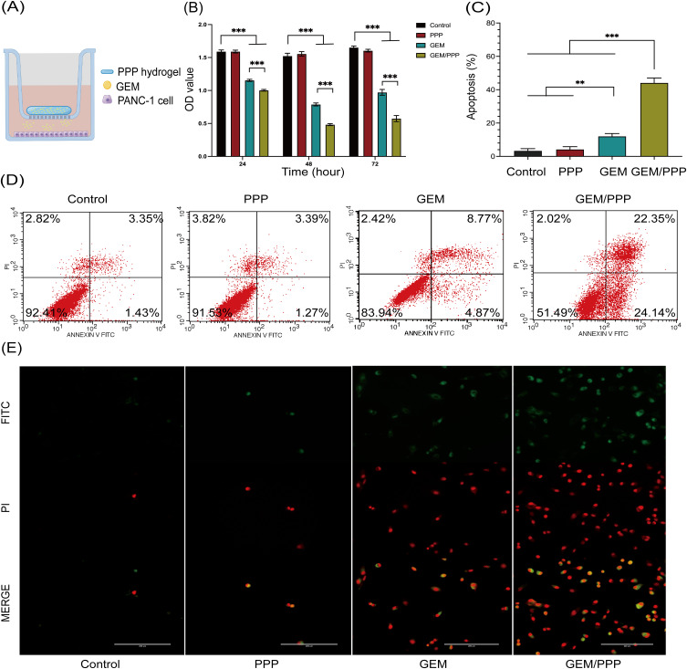
Figure 4.
Cell viability and apoptosis of PANC-1 cells in vitro. (A) Schematic illustration of the Transwell co-culture system used to create a drug depot. (B) Cell viability of PANC-I cells incubated in the Transwell co-culture system was evaluated at 24, 48, and 72 hours. (C) Cell apoptosis of PANC-I cells incubated in the Transwell co-culture system was evaluated at 48 hours. (D) Cell apoptosis analysis measured by flow cytometry (stained by Annexin V-FITC/PI). (E) Representative fluorescent morphology images of the apoptotic PANC-1 cells were obtained after 48 hours of co-cultivation. Double-stained cells represent apoptotic cells. Scale bar is 200 μm. All data were obtained through three independent repeated experiments and presented as the mean ± SD. A significant difference was observed in the LSD post-hoc test. ***P < 0.001, **P < 0.01.

