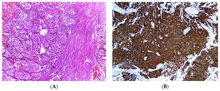
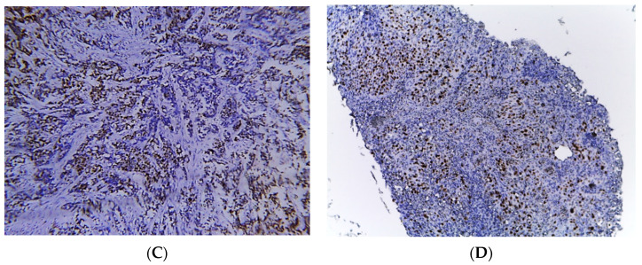
Figure 2.
Photomicrographs of MIBC cases. Histopathological features of muscle-invasive urothelial carcinoma (A). Immunostaining for p16 in MIBC showing strong nuclear and cytoplasmic expression (B). Immunostaining for p53 in MIBC showing positive nuclear expression > 20% of cells (C). Immunostaining for Ki-67 in MIBC showing positive nuclear expression (D) (original magnification ×100).


