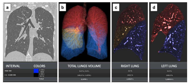
Figure 1.
The Parenchyma Analysis Review step offers a tool to measure the volume between different HU ranges. This protocol provides a measurement/workflow tool that can aid for the assessment of lung diseases. User selectable thresholds can be set to define possible abnormal ranges for the lung parenchyma The regions thus found are visually displayed in the images with different colours (blue colour is for emphysema [not present]; red represent the upper lobes; and yellow the middle lobe) (a). Their measurements are captured and presented in statistics panel including measurements of total lung volume (b) and individual lung volumes (c,d).

