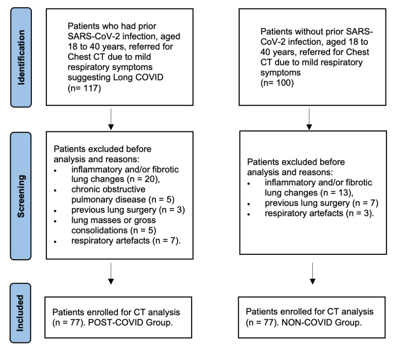
Figure 2.
Study flow diagram of patient recruitment. The initial search of cases identified 117 patients with prior SARS-CoV-2 infection who underwent chest CT due to Long COVID symptoms, of which 40 were excluded for the reasons presented in the diagram. Eventually, 77 cases with prior SARS-CoV-2 infection with no radiologically detectable lung parenchymal abnormalities at chest CT were enrolled (Post-COVID Group). As for control group, 100 patients were initially identified, of which 20 were excluded for the reasons presented in the diagram. Eventually, 77 sex- and age-matched patients were selected as controls (Non-COVID Group).

