Figure 1.
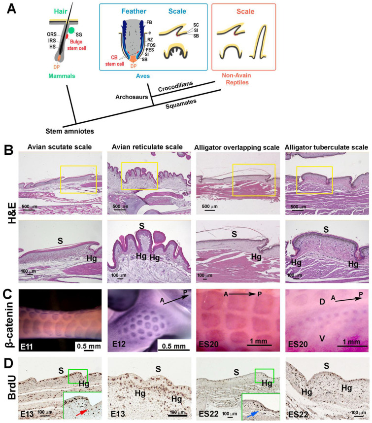
Development of amniote scales: avian scutate scales, avian reticulate scales, reptilian overlapping scales, and reptilian tuberculate scales. (A) Schematic drawing of the architecture of stem cells and niches in amniote skin appendages (modified from [8]). (B) H&E staining showing the chicken scutate scale, reticulate scale, alligator overlapping scale, and alligator tuberculate scale in hatchling chickens and alligators. (C) β-catenin whole-mount in situ hybridization showing the scale primordia. (D) BrdU staining showing the distribution of proliferation cells in developing scales. Note the hinge region has fewer proliferation cells in chicken scutate scales (indicated by the red arrow) whereas the hinge and outer surface regions have similar cell proliferation patterns in chicken reticulate scales and alligator tuberculate scales. There are condensed BrdU-positive cells in the hinge dermal cells of ES22 alligator developing overlapping scales (blue arrow). CB, collar bulge; DP, dermal papilla; e, epidermis; FB; feather barb ridge; FES, feather sheath; FOS, feather follicle sheath; HS, hair shaft; IRS, inner root sheath; M, dorsal middle line of alligator embryo; ORS, outer root sheath; RZ, ramogenic zone; SG, sebaceous gland; SB, stratum basal; SC, stratum corneum; SI, stratum intermedium. A, anterior; D, dorsal; Hg, hinge; P, posterior; S, surface; V, ventral.

