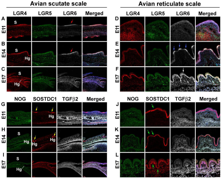
Figure 2.
RNAscope analysis revealing differences in morphogen expression between avian scutate scales and reticulate scales. (A–L) RNAscope assay was used to detect specified RNA transcripts at embryonic day 11, 14, and 17. (A–F) LGR4, LGR5, and LGR6 staining in avian scutate (A–C) and reticulate scales (D–F). The fourth column shows the merged image including DAPI staining. Red arrows in A and B indicate LGR6 expression in the surface of scutate scales. Blue arrows in E indicate LGR6 expression in the surface of reticulate scales. White arrows in (F) show the wide LGR6 expression in reticulate scale epidermis. (G–L) NOG, SOSTDC1, and TGFβ2 staining in avian scutate (G–I) and reticulate scales (J–L). The fourth column shows the merged image including DAPI staining. Yellow arrows in G and H indicate SOSTDC1 expression in the scutate scale hinge area. Green arrows in (J–L) show the wide SOSTDC1 distribution in reticulate scale epidermis. Hg; hinge; S, surface.

