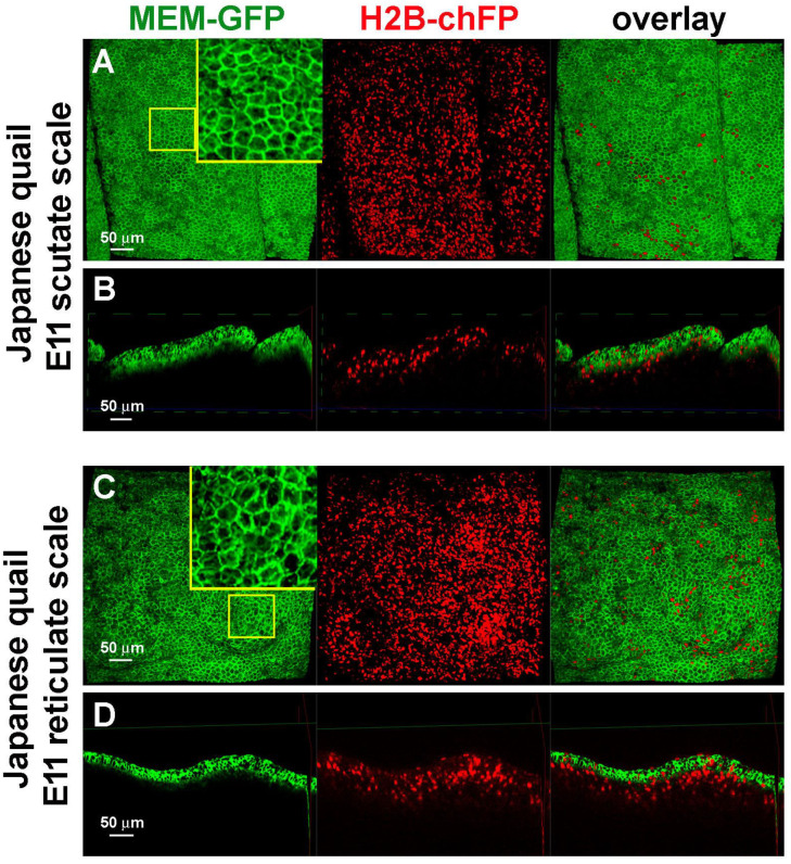
Figure 3.
Confocal 3D images showing the development of scutate scale and reticulate scales using transgenic Japanese quail embryos. (A) Top view of scutate scale primordia at E11. Green expression of membrane-bound GFP and red expression of histone-bound Cherry. (B) Virtual transverse section view of scutate scale primordia at E11. (C) Top view of reticulate scale primordia at E11. (D) Virtual transverse section view of reticulate scale primordia at E11.

