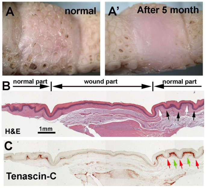
Figure 6.
After full-thickness wounding, avian foot sole repairs the injury but does not regenerate reticulate scales. (A) Bright view of footpad reticulate scales before wounding and (A’) 5 months after wound healing. (B) H&E staining shows the structural differences between normal reticulate scales and the lack of scales within the wound healing region. (C) Tenascin-C immunostaining shows the wound region lacks differential dermal Tenascin-C expression between the normal hinge and surface. Black and white arrows indicate the normal reticulate scale hinge and surface, respectively. Red and green arrows indicate the reticulate scale dermis Tenascin-C expression in the hinge and surface regions.

