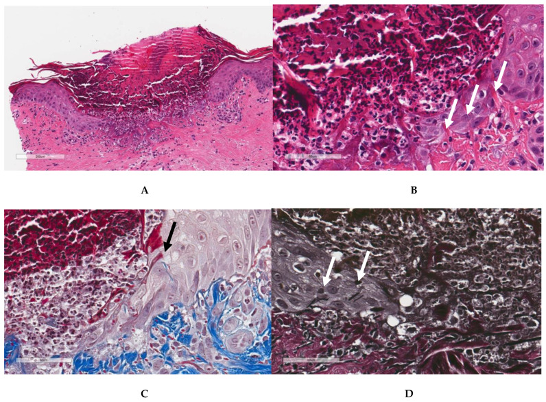
Figure A1.
Reactive perforating collagenosis. (A) Hematoxylin and eosin-stained skin at low magnification shows a cup-shaped depression with degenerating collagen extending from the ulcer base into the crust. (B) High magnification showing the TE of collagen fibers between keratinocytes at one edge of the ulcer (white arrows). (C) Masson’s trichrome confirms the presence of collagen fibers extruding between keratinocytes (black arrow). (D) Verhoeff elastic stain showing extrusion of elastic fragments between keratinocytes at one edge of the ulcer (white arrows).

