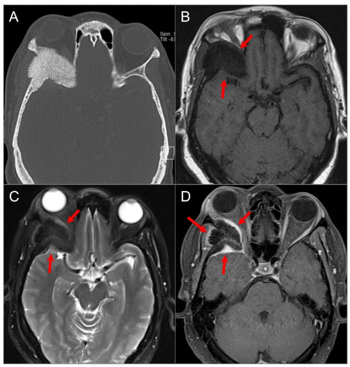
Figure 2.
Intraosseous meningioma. (A) Axial CT shows a marked osseous expansion of the right posterolateral orbital wall and a greater wing of the sphenoid with a spiculated appearance. (B) Axial T1W and (C) STIR T2W images show marked hypointensity in the lesion with thin surrounding soft tissue components along the lateral orbital wall and temporal fossa (arrows). (D) Axial fat-suppressed postcontrast T1W image demonstrates heterogenous transosseous enhancement and avid contrast enhancement in the soft tissue components of the lesion along the temporal dura and orbital/temporal periosteum (red arrows). There is associated displacement of extraocular muscles and proptosis.

