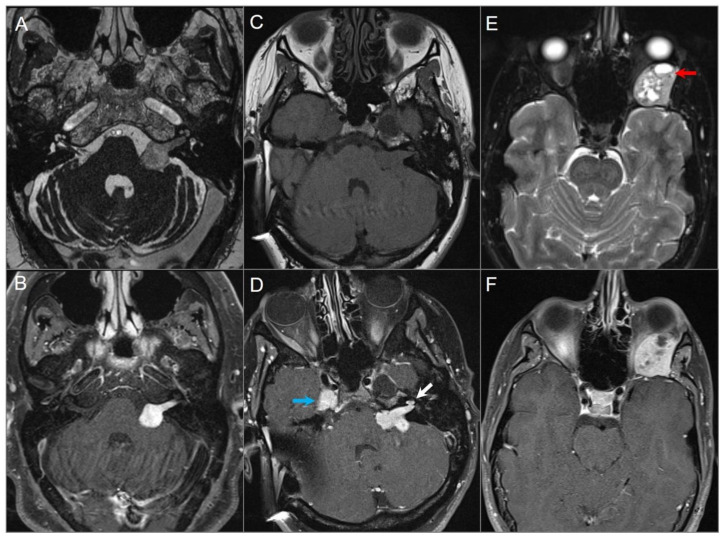
Figure 3.
Schwannomas in three different patients. (A) Axial CISS and (B) axial postcontrast fat-suppressed T1W images show a well-defined, homogenously enhancing left CPA mass with extension into the left internal auditory canal. (C) Axial T1W and (D) postcontrast axial fat-suppressed T1W images show multiple schwannomas centered in the right Meckel’s cave (blue arrow) and the left CPA in a patients with NF2-related schwannomatosis. The left CPA mass completely fills the IAC and extends into the cochlea (white arrow). (E) Axial T2W and (F) axial postcontrast fat-suppressed T1W images reveal a left orbital extraconal, well-defined, heterogeneously enhancing intra-orbital schwannoma with bone remodeling/dehiscence and mass effect on the optic nerve and extraocular muscles. The mass contains multiple non enhancing areas of cystic degeneration with fluid–fluid levels likely due to hemorrhage (red arrow).

