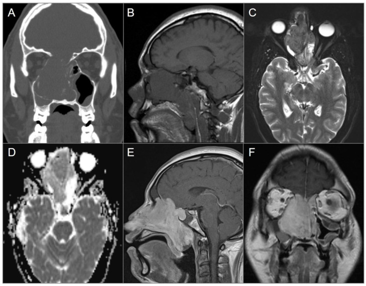
Figure 10.
Nasal osteosarcoma invading skull base. (A) Coronal CT image shows a bulky expansile soft tissue density lesion in the nasal cavity, eroding the anterior skull base, nasal septum, and right lamina papyracea. (B) Sagittal T1W and (C) axial fat-saturated T2W MR images reveal slightly hypointense signal of the lesion on both sequences as well as mucus accumulation in the adjacent paranasal sinuses due to obstruction of the drainage pathways. (D) ADC map reveals mild diffusion restriction in the lesion anteriorly. (E) Sagittal and (F) coronal postcontrast T1W images demonstrate avid enhancement of the lesion with extension to the anterior cranial fossa, nasopharynx, and oropharynx.

