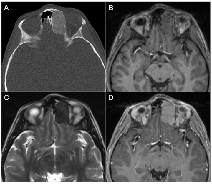
Figure 11.
Ossifying fibroma. (A) Axial CT image shows an expansile and well-defined ground-glass density lesion with a thin sclerotic rim, centered in the left frontoethmoid sinuses. Note remodeling/scalloping of the left medial orbital wall and bulging into the left anterior cranial fossa. (B) T1W and (C) T2W axial images reveal isointense signal on T1 and markedly hypointense signal on T2. (D) Axial postcontrast T1W image demonstrates moderate homogenous enhancement of the lesion.

