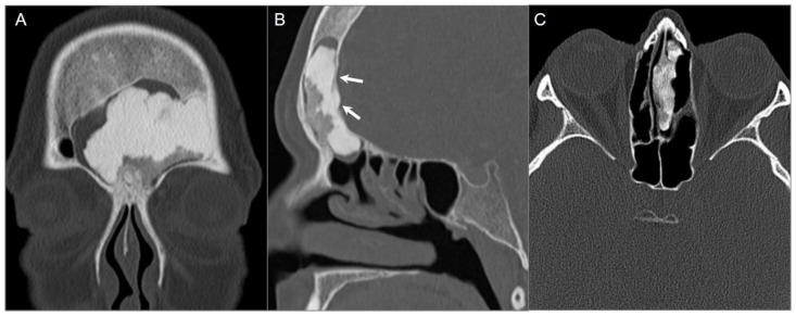
Figure 12.
Osteomas in two different patients. (A) Coronal and (B) sagittal CT images show a large frontal sinus osteoma with mixed dense and hypodense areas consistent with cortical and cancellous components, respectively. Note mild intracranial extension (arrows). (C) Axial CT image in a different patient demonstrates a well-defined, heterogeneously hyperdense lesion centered in the left ethmoid sinus and attached to the nasal septum.

