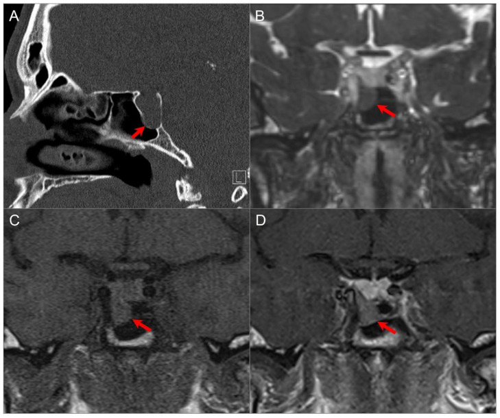
Figure 14.
Exophytic/ectopic-intrasphenoidal pituitary adenoma. (A) Sagittal CT image shows erosion of the sellar floor with a soft tissue lesion extending into the clivus and posterior margin of the sphenoid sinus (red arrow). (B) Coronal postcontrast CISS and (C) noncontrast T1W images reveal a hypointense lesion abutting the inferior margin of the right pituitary lobe and medial wall of the petrocavernous ICA. (D) Coronal postcontrast T1-weighted image demonstrates normal enhancement of the pituitary gland and hypoenhancement of the exophytic adenoma (red arrow).

