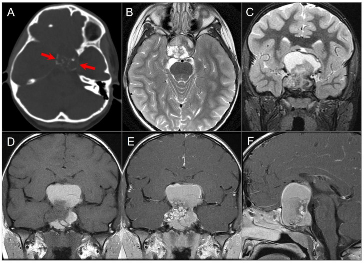
Figure 16.
Adamantinomatous craniopharyngioma in a child. (A) CT image shows a partially calcified sellar and suprasellar mass (red arrows). (B) Axial and (C) coronal T2W, (D) coronal noncontrast T1W, and (E) coronal and (F) sagittal postcontrast T1W images show a complex mass with mixed cystic and solid components. Note: hyperintense T1 and T2 signal in the cystic components consistent with proteinaceous, hemorrhagic and/or cholesterol contents. The mass demonstrates intrasphenoidal and intraclival extension with mild prepontine bulge and suprasellar extension with mass effect on the optic chiasm and third ventricle. Heterogenous enhancement of the solid tumor components is noted.

