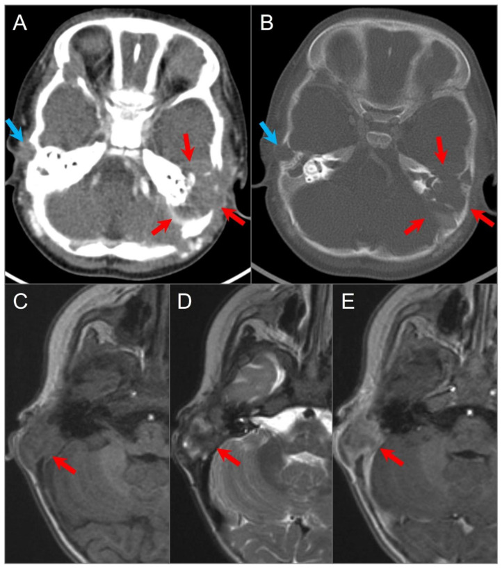
Figure 17.
Langerhans cell histiocytosis in a 9-month-old boy. Axial postcontrast CT in soft tissue (A) and bone window (B) settings shows a lytic ‘punched out’ bone lesion involving the left petromastoid temporal bone with middle and posterior cranial fossa extensions (red arrows). The lesion also erodes the left semicircular canals and involves the left middle ear cavity and facial nerve. Another small destructive lesion with similar CT characteristics is seen in the right temporal bone (blue arrows). In a different 3-year-old boy, (C) noncontrast T1W, (D) T2W, and (E) postcontrast T1W axial images demonstrate a relatively well-delineated lesion in the right temporal bone (red arrows). The lesion demonstrates a slightly heterogeneous isointense signal on T1 and mixed hypointense and hyperintense signals on T2WI with heterogeneous enhancement after gadolinium administration.

