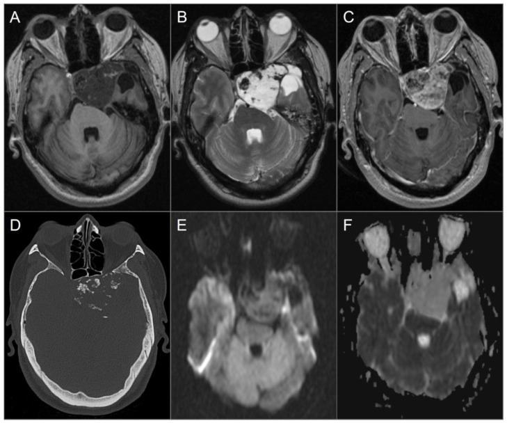
Figure 20.
Chondrosarcoma. (A) Axial noncontrast T1W and (B) T2W images show a well-circumscribed T1 hypointense, markedly T2 hyperintense lesion centered in the left petroclival fissure involving the clivus, cavernous sinus, posterior aspect of the sphenoid sinus and left middle cranial fossa. The lesion extends into the prepontine cistern and compresses the brain stem, left temporal lobe, and basilar artery. (C) Axial postcontrast T1W image shows avid heterogeneous enhancement of the mass. (D) Axial CT image shows permeative destructive margins and chondroid calcifications in the lesion. (E) DWI and (F) ADC map images reveal facilitated diffusion in the lesion.

