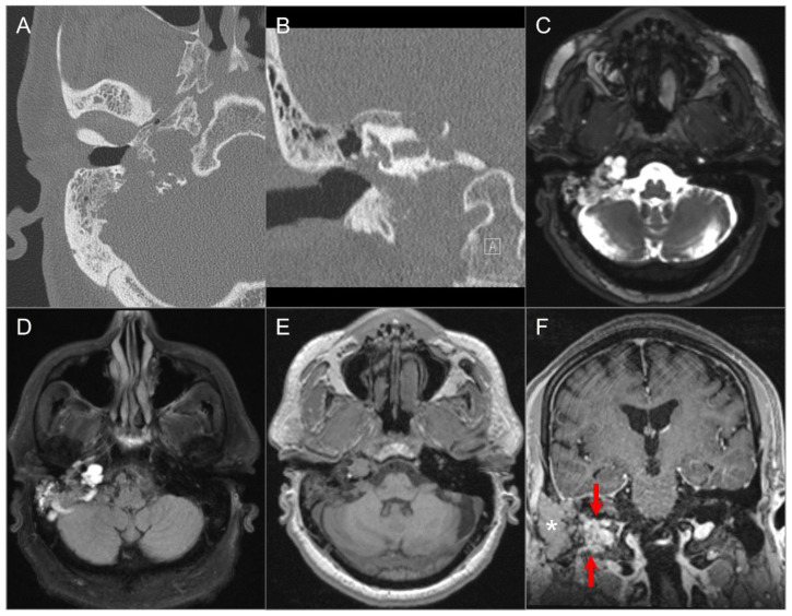
Figure 22.
Endolymphatic sac tumor. (A) Axial and (B) coronal CT images show a lytic lesion with a moth-eaten, permeative appearance in the right retro-labyrinthine petrous temporal bone extending to the middle ear cavity, mastoid region, and jugular fossa. (C) Axial fat-suppressed T2W, (D) axial FLAIR, and (E) axial noncontrast T1W images demonstrate a complex solid and cystic lesion with mixed signal intensity, likely reflecting hemorrhagic and proteinaceous contents. (F) Postoperative coronal postcontrast T1-weighted image shows heterogeneous enhancement in the residual solid component of the lesion (red arrows). The more lateral tissue corresponds to a fat graft (asterisk).

