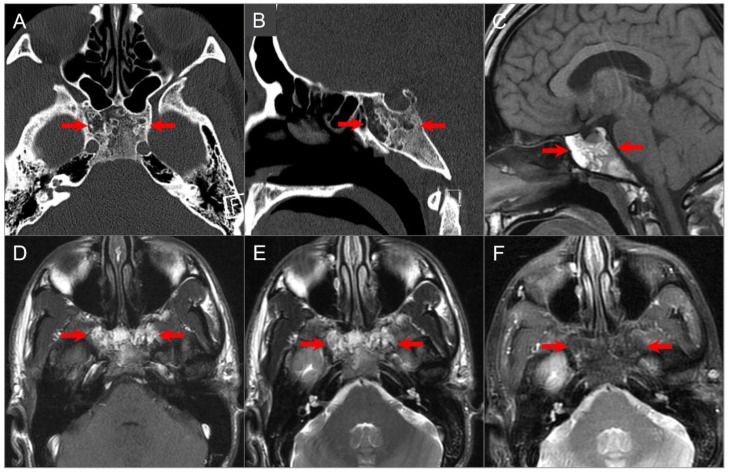
Figure 24.
Arrested pneumatization. (A) Axial and (B) sagittal CT images show partial pneumatization of the sphenoid sinuses anteriorly with a heterogeneous appearance of the sphenoid body. There are multiple hypodense foci with peripheral sclerosis, a non-expansile and non-destructive appearance, and similar density to subcutaneous fat (red arrows). (C) Sagittal and (D) axial noncontrast T1W and (E) axial T2W images reveal heterogeneous hyperintense signals in these regions. (F) Fat-suppressed T2W image shows signal suppression consistent with fatty bone marrow.

