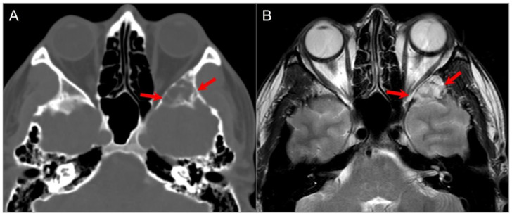
Figure 25.
Sphenoid wing cephalocele. (A) Axial CT image reveals a lobulated, well-defined lucent lesion in the left greater sphenoid wing with bony scalloping and thin internal septations (red arrows). (B) T2W axial MR image shows CSF isointense signal within the lesion with linear hypointense septations.

