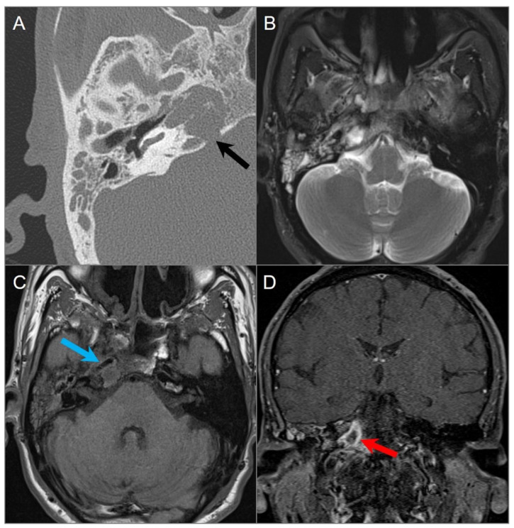
Figure 30.
Petrous apicitis. (A) Temporal bone CT image shows opacification of the right mastoid air cells and external auditory canal with thickening of the tympanic membrane consistent with external otitis and mastoiditis. There is opacification of the right petrous apex with dehiscence of its posteromedial margin (black arrow). (B) Axial T2W and (C) noncontrast T1W images demonstrate fluid in the mastoid air cells, middle ear cavity, and petrous apex with associated thickening of the carotid artery wall, resulting in mild luminal narrowing (blue arrow). (D) Coronal postcontrast fat-suppressed T1W image reveals a peripherally enhancing collection within the right petrous apex consistent with abscess (red arrow).

