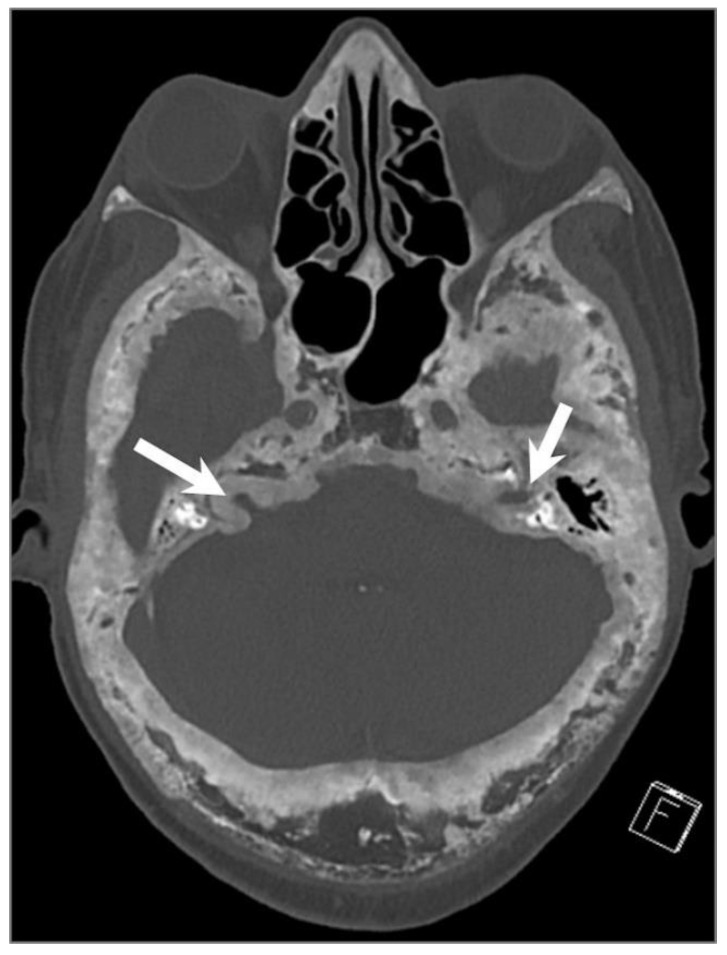
Figure 31.
Paget’s disease. Axial CT image shows characteristic mixed lytic and sclerotic areas (‘cotton wool’ appearance) with thickened trabeculae, bone expansion, cortical thickening, and deformity of the calvarium and skull base. There is narrowing of the bilateral internal auditory canals due to cortical thickening (arrows).

