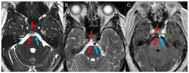
Figure 32.
Ecchordosis physaliphora. (A) Axial CISS, (B) axial T2W, and (C) axial postcontrast T1W images reveal a small, lobulated, and markedly T2 hyperintense lesion in the clivus. There is no associated contrast enhancement. The lesion protrudes into the prepontine cistern (red arrows) abutting the ventral pons and basilar artery (blue arrows).

