Figure 1.
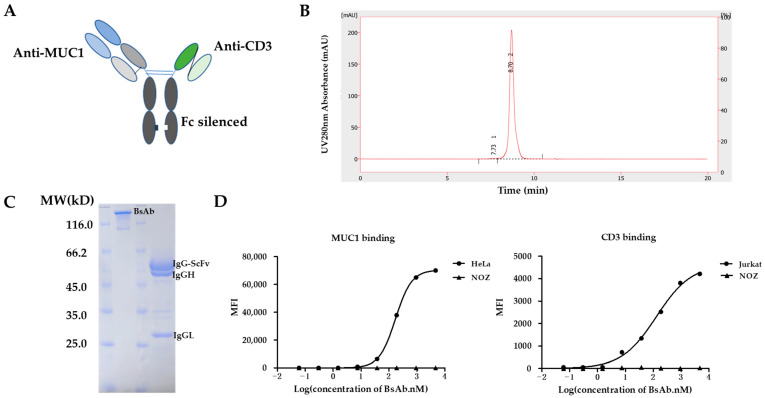
Generation of MUC1/CD3 BsAb. (A) Schematic design of MUC1/CD3 BsAb. (B) SEC-HPLC analysis of the purified MUC1/CD3 BsAb. The percentage of the area of peak 1 and peak 2 is 0.7% and 99.3% respectively, which represents the high purity of the purified BsAb. (C) SDS/PAGE and Coomassie blue staining results of the purified MUC1/CD3 BsAb under non-reducing and reducing conditions. (D) Cell binding analysis of MUC1/CD3 BsAb to MUC1 and CD3-positive cells by flow cytometry, HeLa (MUC1-positive cells), Jurkat (CD3-positive cells), NOZ (CD3 and MUC1-negative cells).

