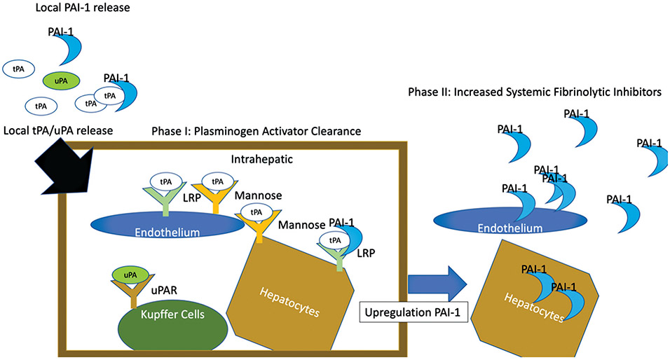
Fig. 4.
Schematic of the two phases of fibrinolysis shutdown. This figure represents a schematic of the proposed mechanism of fibrinolysis shutdown. Plasminogen activators released in circulating blood are initially controlled by local PAI-1 levels. Activators surpass the local PAI-1 binding and enter the liver. The numerous receptors of cells in the liver that have tPA avidity bind the plasminogen activators. This is the phase I of fibrinolysis shutdown. Following this, the response to tPA from local liver cells increases PAI-1 synthesis, resulting in increased release of PAI-1 up to an hour after activator clearance. Additional peripheral endothelial cells also play a role in increase PAI-1 production from cytokine release. This second phase of shutdown is hallmarked by resistance to fibrinolytic activators. PAI-1, plasminogen activator inhibitor 1; tPA, tissue plasminogen activator; uPA, urokinase-type plasminogen activator.

