
Figure 1.
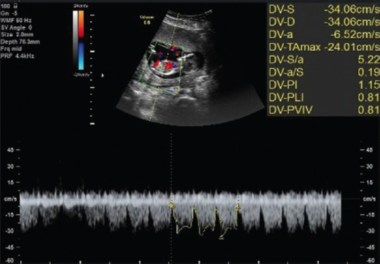
The color and pulsed Doppler ultrasonographic visualization of DV. A mid-sagittal view of the fetal trunk shows DV in the distal portion of the umbilical vein (upper part). Three typical DV waveforms were manually traced (lower part); DVPI, DVPLI (equivalent to DVRI), and DV S/A were extracted (upper right part). DV = Ductus venosus, DVPI = DV pulsatility index, DVPLI = DV preload index, DVRI = DV resistance index