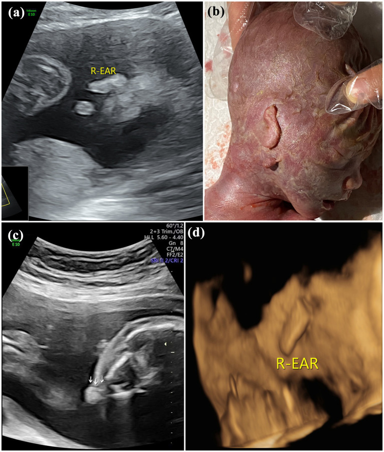
Figure 3.
Images of type III of microtia: (A) 2D sonography and (B) photo (induced labor) of microtia type III accompanied by an accessory auricle; (C) 2D sonography of unilateral microtia type III accompanied by atresia of external acoustic meatus; and (D) 3D sonography of Unilateral microtia type III.

