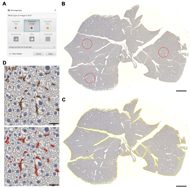
Figure 3. Macrophage detection using LiverQuant.
A) Upon opening the chosen DAB-stained image, set the image type to Brightfield H-DAB. B) Example of three annotations used for the Macrophage intensity script to gather an average intensity of DAB staining. C) The Macrophage detection script will automate tissue annotation and D) generate positive macrophage detections (red). B and C, scale bars: 2 mm. D, scale bars: 25 μm.

