
Figure 1.
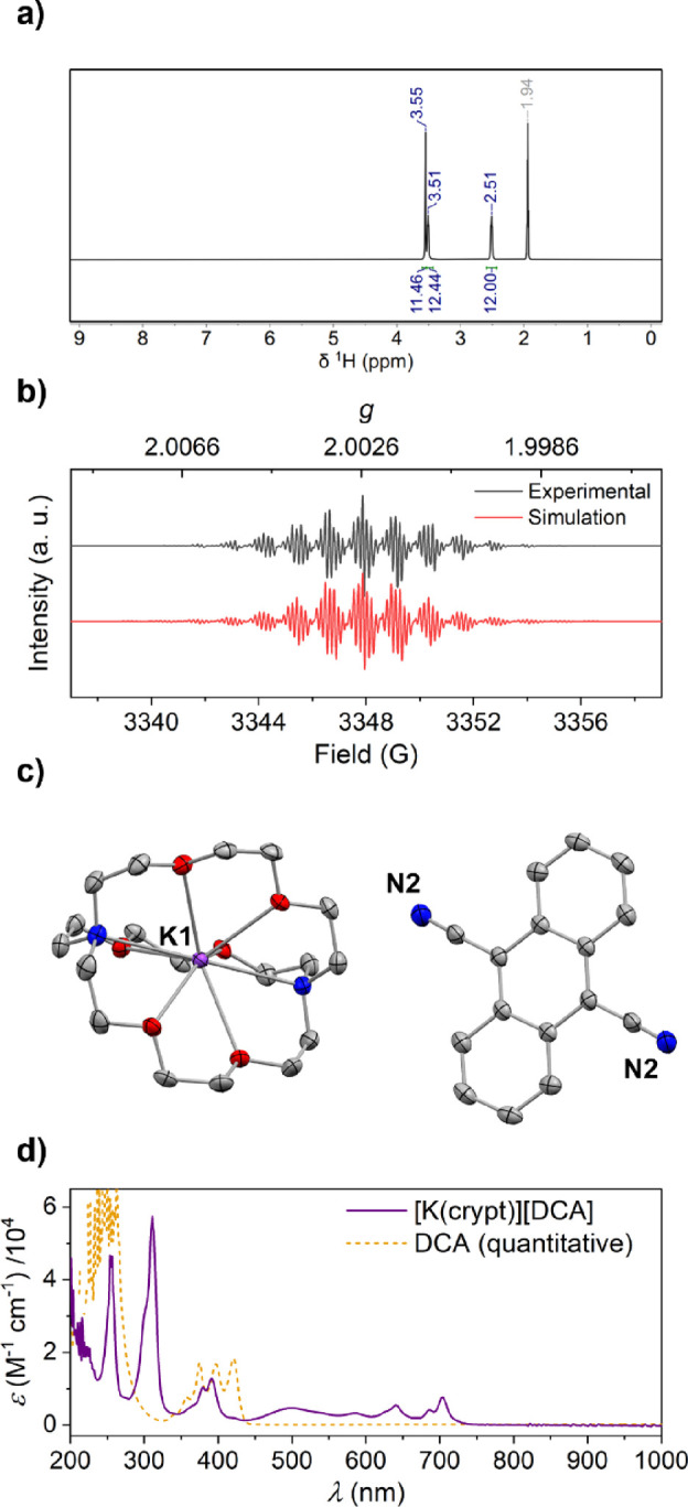
Spectroscopic and crystallographic characterization of [K(crypt)+][DCA·–] (additional details in the SI): (a) 1H NMR spectrum (CD3CN (δ = 1.94 ppm), RT); (b) X-band EPR spectrum (4:1 PhMe/THF, 109 μM, RT; simulated as g = 2.00256, 4 × A(1H) = 3.904 MHz, 4 × A(1H) = 2.969 MHz, 2 × A(14N) = 0.4307 MHz, 15% A(13C) = 20.631 MHz); (c) single-crystal XRD structure; thermal ellipsoids shown at 50%; H atoms omitted for clarity and only one of two inequivalent DCA moieties shown (asymmetric unit contains 1 × [K(crypt)+] and 2 × 0.5[DCA·–]); C atoms in gray, N in blue, O in red, K in purple; (d) UV–vis spectrum (MeCN, 544 μM, 1 mm path, RT).