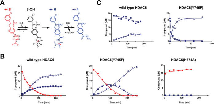
Figure 3.
Inhibitor hydrolysis by purified HDAC6 variants in vitro. Panel (A): Hydrolysis scheme of 8 by HDAC6. Cleaved bonds are marked by red lines. Panel (B): Kinetics of compound 8 hydrolysis by wild-type HDAC6 and HDAC6(Y745F) and HDAC6(H574A) mutants. Compound 8 (20 μM final concentration) was mixed with 2 μM wild-type HDAC6 in the assay buffer, and reaction mixtures were incubated at 30 °C. Aliquots of reaction mixtures were analyzed by LC–MS/MS at given time points by quantifying concentrations of compounds 4, 6, and 8. Analysis of a putative intermediate 8-OH was not technically possible as it is unstable in aqueous solutions and thus cannot be synthesized to generate a corresponding analytical method and calibration curve. No hydrolysis of compound 8 was observed for the HDAC6(H574A) mutant. Panel (C): Kinetics of compound 6 hydrolysis by wild-type HDAC6 and the HDAC6(Y745F) mutant. Reaction conditions and quantifications were identical as described for panel (B).

