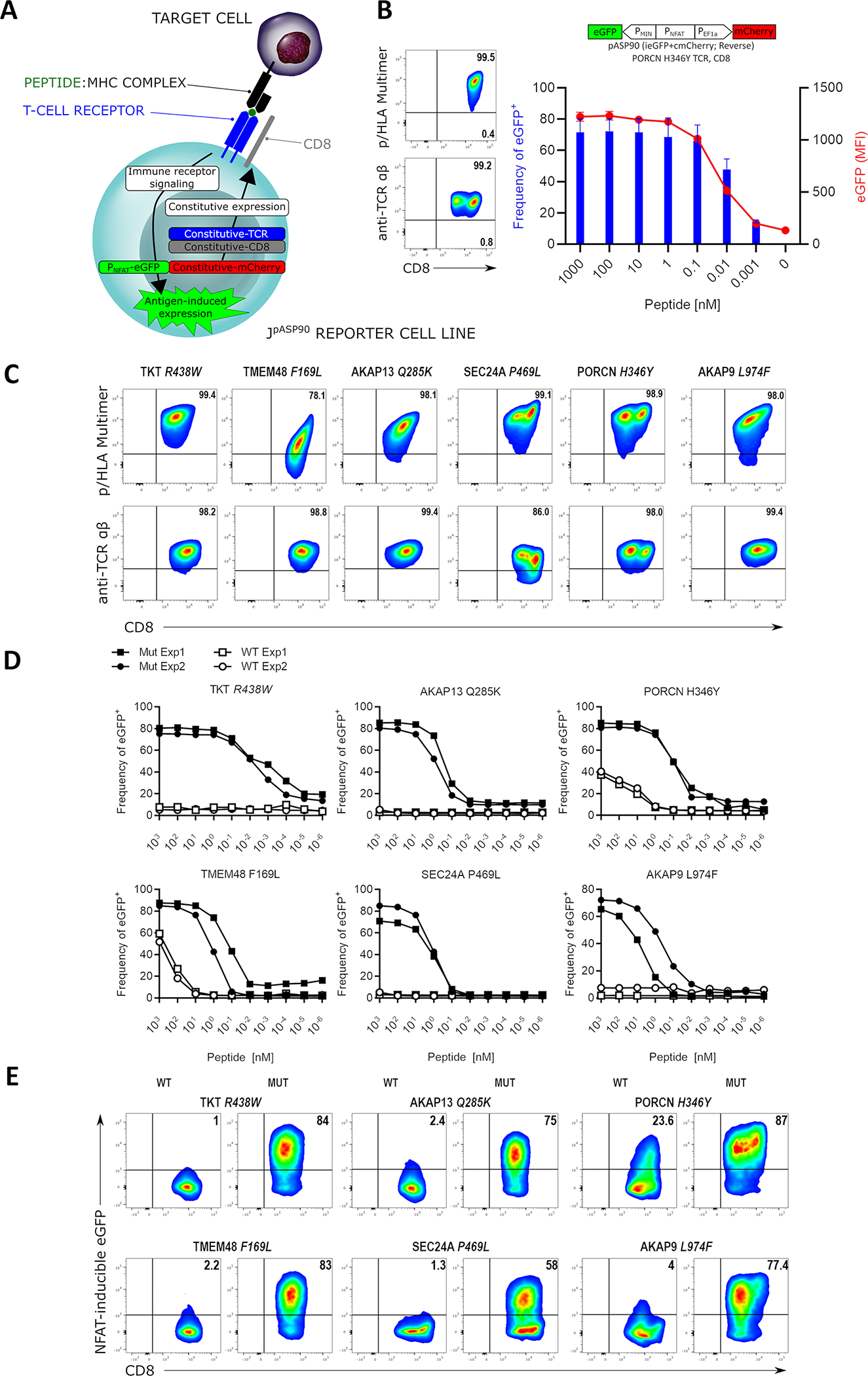
Figure 3: Uni-Vect Expression Cassettes for Characterization of T-cell Receptors.

(A) Schematic representation of Uni-Vect-Jurkat reporter cell lines JpASP90 and JpASP89. (B) Transduction of TCR constructs into JpASP90 reporter cell lines. Titrated concentrations of peptide (LLHGFSFYL) antigen in the presence of antigen presenting cells led to upregulation of NFAT-inducible eGFP expression as a surrogate for activation via TCR signaling. Blue bars represent percentage of eGFP positive cells, red line represents MFI of eGFP at each concentration. (n=3). (C) Flow cytometry analysis of 6 neoantigen-specific/HLA-A*02:01 restricted TCRs engineered into the JpASP90 reporter cell line. (D) NFAT reporter EC50 curves for neoantigen-specific TCRs expressed in JpASP90 reporter cell line upon activation with mutated (blue) and wild-type (red) peptides. (E) JpASP90 reporter cell line expressing the various TCRs were cultured for 16 h with melanoma cell line DM6 expressing tandem mini-gene constructs encoding mutated or wild-type antigen and analyzed for inducible eGFP expression. All data are representative of two or more experiments. All data with error bars are presented as mean ± SD. See also Figure S4 and Tables S1 and S2.
