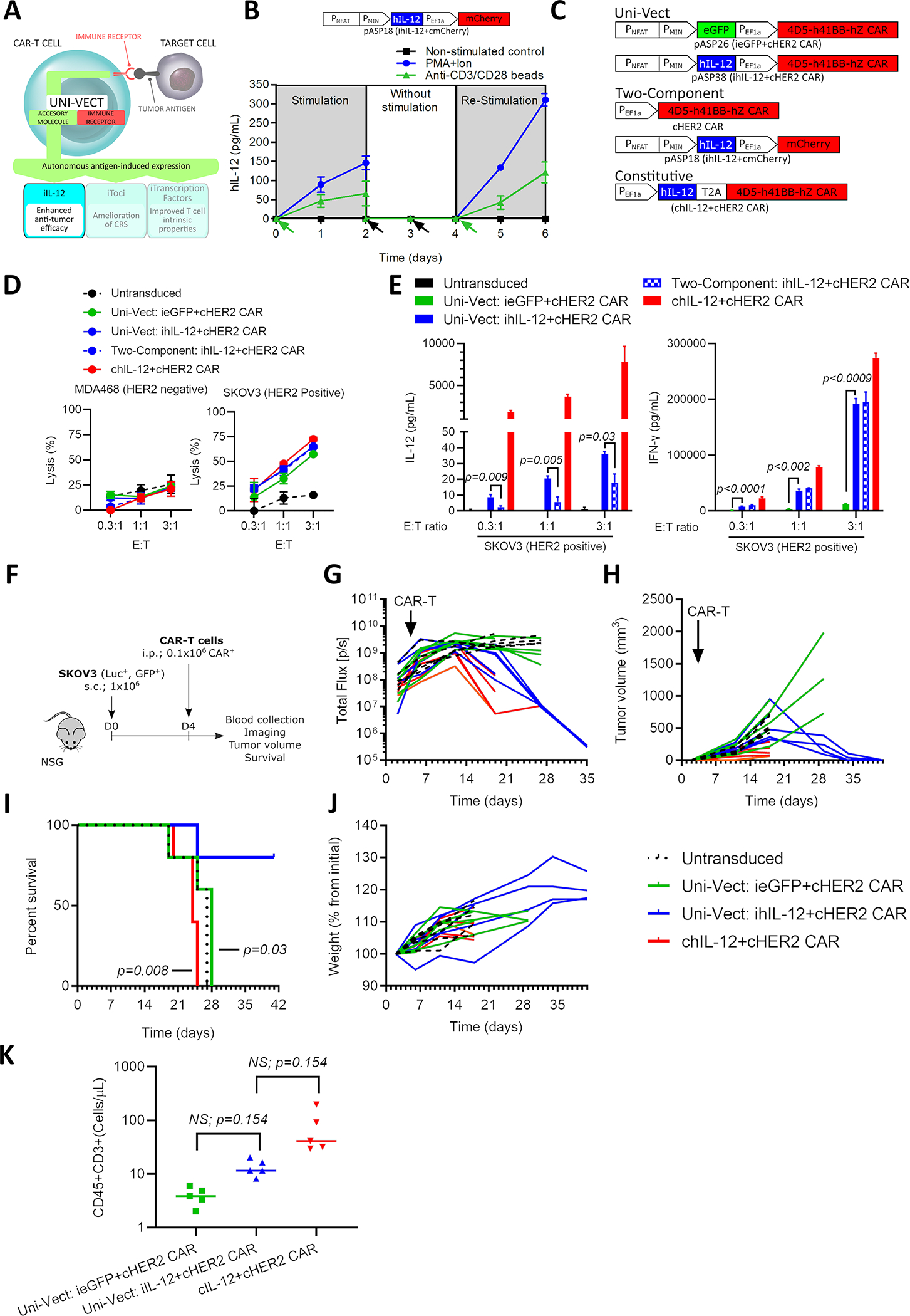
Figure 4: iIL-12 Enabled Uni-Vect Improves Antitumor Efficacy of CAR-T Cells In Vivo.

(A) Schematics of iIL-12 integrated into Uni-Vect. (B) Primary T cells transduced with pASP18 were stimulated for 2 days, followed by 2 days of rest, and then re-stimulated for another 2 days. IL-12 secretion was measured at 24 h increments. (n=3). Green arrow represents stimulation and black arrow represents media exchange. (C) Schematic representation of compared constructs/experimental groups. (D) CAR-T cells were co-cultured with HER2+ and HER2− target cell lines. Lysis was measured by luciferase assay. (n=3) (E) IL-12 and IFN-γ secretion from co-cultures in (D). (Welch ANOVA) (F) In vivo study design. CAR-T cells were injected i.p. at 0.1 × 10⁶ CAR-T per mouse in NSG mice with established HER2+ SKOV3 ovarian cancer tumors. (n = 5 mice per group) (G) Quantified tumor luciferase activity. (H) Tumor volume as measured by caliper. (I) Percent survival in each group (Log-rank Mantel-Cox test). (J) Plot of mouse weight versus time by group (n=5). (K) CD3+ T cell counts in peripheral blood at day 17. (Kruskal-Wallis test). i.p; Intraperitoneal, s.c.; Subcutaneous. Each line in G, H, and J and each dot in K represents individual mice. NS; not significant. All data are representative of two or more experiments. All data with error bars are presented as mean ± SD. See also Figure S5 and Table S1.
