
Fig. 4. Recombinant NTF3 increases hepatocyte CCND1 in HSC-depleted mice.
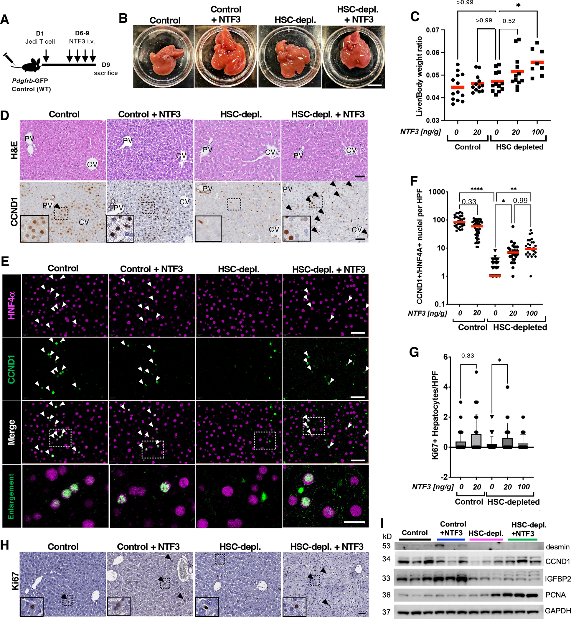
(A) Experimental scheme. (B) Macroscopic images of livers in control and HSC-depleted mice treated with vehicle or NTF3. Scale bar, 1 cm (C) Liver-to-body weight ratios of control and HSC-depleted mice treated with vehicle or the indicated concentrations of NTF3. *p<0.05 by Kruskal-Wallis, post hoc Dunn’s test. n=8–13 animals per group. (D) H&E and CCND1 staining of liver sections from control and HSC-depleted mice treated with vehicle or NTF3. Arrowheads indicate positive staining. Dashed rectangles indicate areas shown in insets. (E) Multiplex immunohistochemistry for CCND1 and HNF4α in liver sections from control and HSC-depleted mice treated with vehicle or NTF3. Dashed rectangles indicate areas of enlargement. Scale bars, 20 μm. Arrowheads indicate positive-staining nuclei. (F) Quantification of CCND1+HNF4α+ nuclei per high power field (HPF). Red bars indicate mean. n=3–5 animals per group. *p<0.05, **p<0.005, ****p<0.0001 by Kruskal-Wallis, post hoc Dunn’s test. (G) Quantification of Ki67+ hepatocytes in control and HSC-depleted mice treated with vehicle or NTF3. Mean+SD. *p<0.05 by Kruskal-Wallis, post hoc Dunn’s test. n=4–6 animals per group. (H) Ki67 staining of liver sections from control and HSC-depleted mice treated with vehicle or NTF3. Arrowheads indicate positive staining. Dashed rectangles indicate areas shown in insets. (I) Western blot for desmin, CCND1, IGFBP2, and PCNA in whole-liver lysates from control and HSC-depleted mice treated with vehicle or recombinant NTF3. GAPDH is a loading control. N=3 animals per group.