Fig. 1.
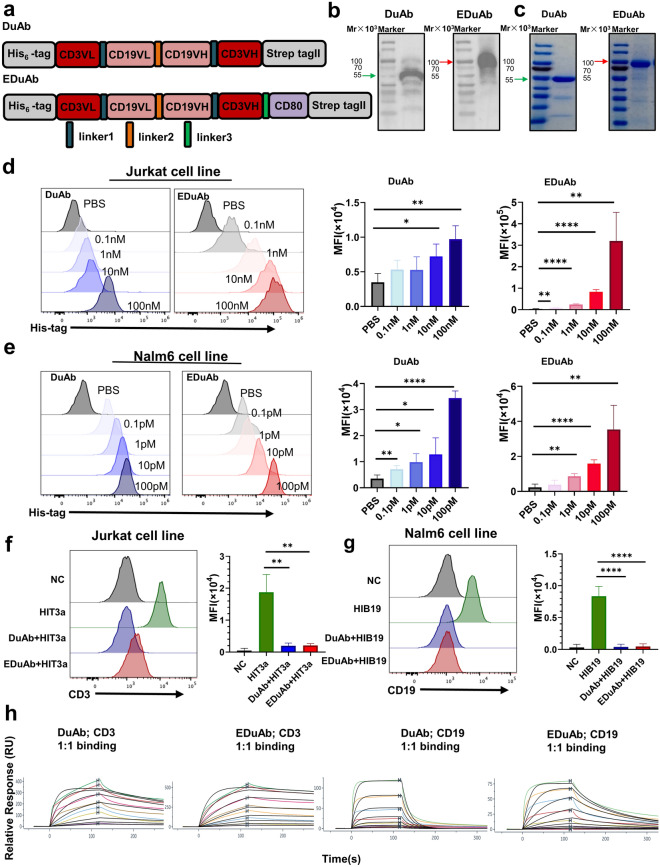
Design, production, and binding specificity of DuAb and EDuAb. a Schematic diagram of DuAb and EDuAb construct. b Western blot analysis of DuAb and EDuAb in supernatant of ExpiCHO-S cells transduced with expression plasmids. c Purified DuAb and EDuAb were analyzed by SDS-PAGE gel. d–e The binding affinity of DuAb and EDuAb to CD3+ and CD19+ cells were determined using flow cytometry (Left panel). MFI quantification and statistical analysis of the data (Right panel). f–g Competitive binding activity of DuAb and EDuAb with commercial PE-CY7-conjugated HIT3a and PE-CY7-conjugated HIB19 were detected using flow cytometry (Left panel). MFI quantification and statistical analysis of the data (Right panel). h The binding kinetics of DuAb or EDuAb to CD3 and CD19 protein were determined by Surface Plasmon Resonance (SPR)

