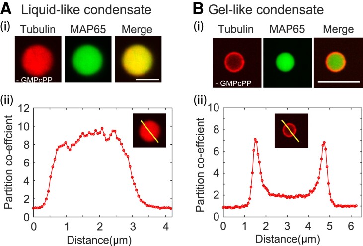
Fig. 3.
Tubulin colocalization and microtubule organization with MAP65 condensates. A) Tubulin co-localizes with MAP65 liquid droplets. (i) Representative confocal image of rhodamine tubulin (red, left) and GFP-MAP65 (green, center) and overlay (merge, right) in a liquid-like droplet. Scale bar is 5 m. (ii) Intensity scan through the droplet in the rhodamine channel normalized so that the intensity outside is one. B) Tubulin co-localizes with MAP65 matured droplets. (i) Representative confocal images of rhodamine tubulin (red, left) and GFP-MAP65 (green, center) and overlay (merge, right) in a gel-like droplet. Scale bar is 5 m. (ii) Intensity scan through the droplet in the rhoamdine channel normalized so that the intensity outside the droplet is one.

