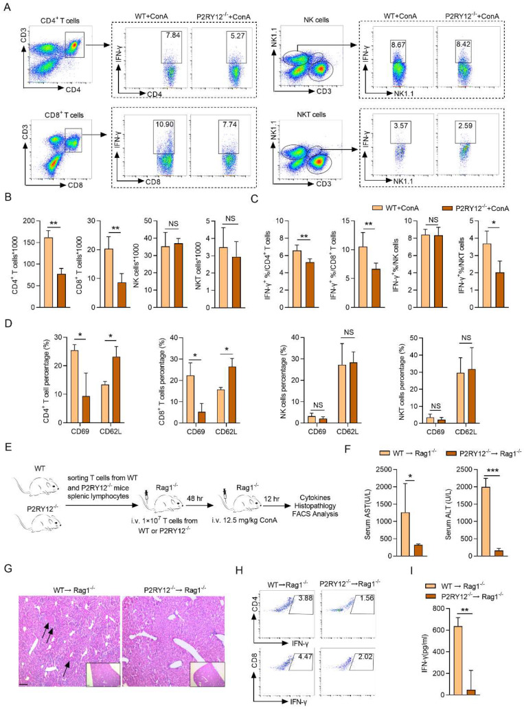
Figure 2.
P2RY12 in T cells exacerbates the pathology of ConA-induced immune hepatitis. WT mice and P2RY12-/- mice were administered ConA (12.5 mg/kg body weight via i.v. injection) for 12 hr. (A) IFN-γ expression in CD4+ T cells, CD8+ T cells, NK cells, and NKT cells from liver lymphocytes of WT mice and P2RY12-/- mice were analyzed by flow cytometry. (B) Total numbers of liver lymphocytes, including CD4+ T cells, CD8+ T cells, NK cells, NKT cells were observed in the liver of WT mice and P2RY12-/- mice. (C) The proportion of IFN-γ+ in CD4+ T cells, CD8+ T cells, NK cells, NKT cells of WT + ConA mice and P2RY12-/- + ConA mice were examined by flow cytometry. (C) Pooled data are presented from (A). (D) The expression of CD62L and CD69 in CD4+ T cells, CD8+ T cells, NK cells, NKT cells in Figure 2A were analyzed by flow cytometry. (E) Schematic representation of adoptive transfer assay of WT and P2RY12-/- mice. T cells purified by splenic lymphocytes from WT mice and P2RY12-/- mice (8-10 weeks, n = 8) were transferred into Rag1-/- mice through tail vein injection, and 48 hr later, these mice were followed 12.5 mg/kg ConA for 12 hr. (F) The serum levels of AST and ALT were detected. (G) Photomicrographs of representative H&E-stained mouse livers. Massive hepatocyte necrosis (dark arrows) was observed in WT→Rag1-/- mice. (H) The proportion of IFN-γ in CD4+ T cells, CD8+ T cells from liver lymphocytes of WT→Rag1-/- mice and P2RY12-/-→Rag1-/- mice were analyzed by flow cytometry. (I) The serum level of IFN-γ was detected. One representative data of three independent experiments was shown. Data are mean ± SEM. *P< 0.05; **P< 0.01; ***P < 0.001 vs indicated group. (two-tailed Student's t-test).

