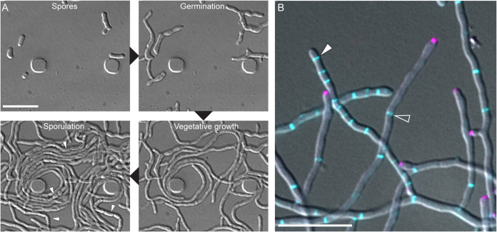
FIG 3.
Streptomyces venezuelae. (A) Time-lapse images of wild-type S. venezuelae grown in liquid using a microfluidic system showing the cellular development during the Streptomyces life cycle, including germination, vegetative growth, and sporulation (white arrowheads). (B) Composite light microscopy image of S. venezuelae producing fluorescently tagged DivIVA (magenta) and FtsZ (blue) to visualize growing hyphae and sites of cell division, respectively. Open arrowheads indicate crosswalls, and filled arrowheads point to hyphae undergoing sporulation septation. Scale bars, 10 μm.

