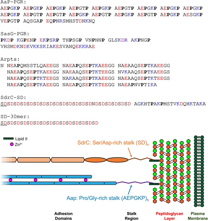
FIGURE 1.

Sequences of IDP constructs. Prolines are in bold font, negatively charged residues are colored red, and positively charged residues are colored blue. Sequence repeats are separated by spaces to highlight the pattern. The illustration at the bottom highlights the location of the stalk regions relative to the peptidoglycan layer.
