
FIGURE 2.
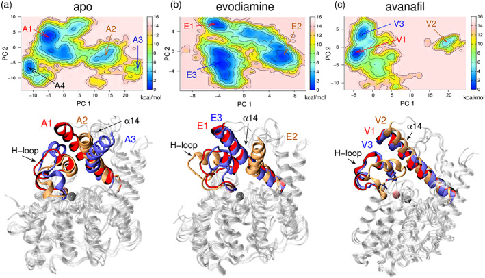
The free energy surface and the structures corresponding to the metastable states. (Top panel) The free energy surface and the metastable states are labeled for each of the three PDE5 states (apo, evodiamine‐bound, and avanafil‐bound). (Lower panel) The representative structures of PDE5 corresponding to the metastable states are shown in cartoon representations. The conformations of the H‐loop and the α14 helix in each metastable state are uniquely colored and shown in cartoon representations. In the free energy surface of the PDE5‐apo form (a), four metastable states are labeled: A1 (red), A2 (orange), A3 (blue), and A4 (black). The PDE5 structure corresponding to the A1 (red), A2 (orange), and A3 (blue) states are shown, and the structure of the A4‐state is similar to the A1‐state (see Figure S4D). In the free energy surface of the PDE5‐evodiamine form (b), three metastable states are shown: E1 (red), E2 (orange), and E3 (blue). In the free energy surface of PDE5‐avanafil form (c), three metastable states labeled V1 (red), V2 (orange), and V3 (blue), are shown. The PDE5 structure of the V3‐state is similar to the V1‐state. The zinc (gray) and the magnesium (pink) ions, located in the catalytic site, are shown as spheres.