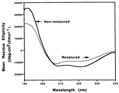
FIG. 9.
Secondary structure analysis of nonrenatured and renatured rTromp1 by CD spectroscopy. The double minimum absorbances displayed by nonrenatured rTromp1 (solid line) is typical of proteins with a high percentage of alpha-helix. By comparison, renatured rTromp1 (dotted line) showed a distinct loss in absorbance at 222 nm, indicating a loss in alpha-helical secondary structure.

