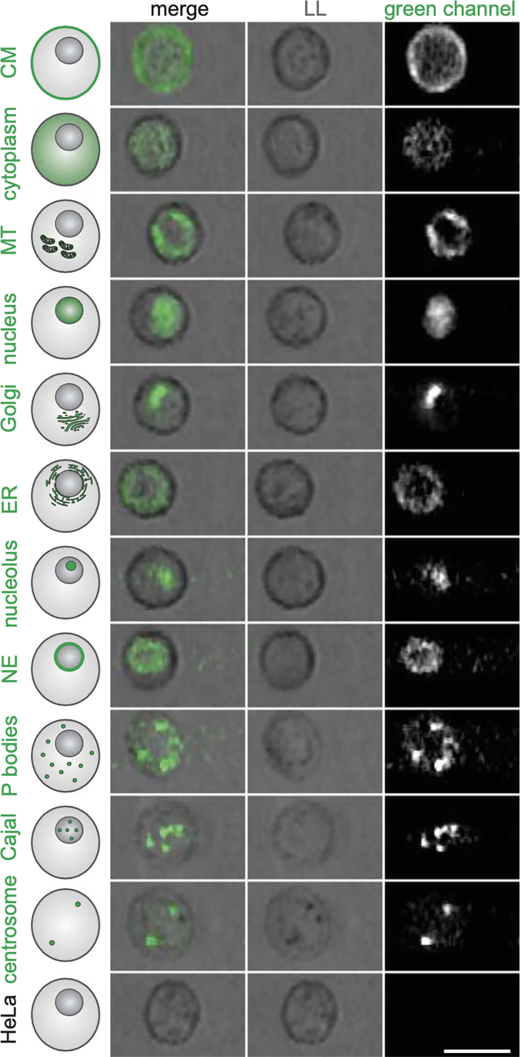
Figure 1. Image-Based Localization Phenotypes Detectable in Cells in Suspension.

A variety of protein localizations can be detected, classified, and sorted with the use of an image-based cell sorter. Light loss imaging is similar to traditional light field microscopy. (Adapted from Schraivogel et al.1 with permission from the American Association for the Advancement of Science.)
