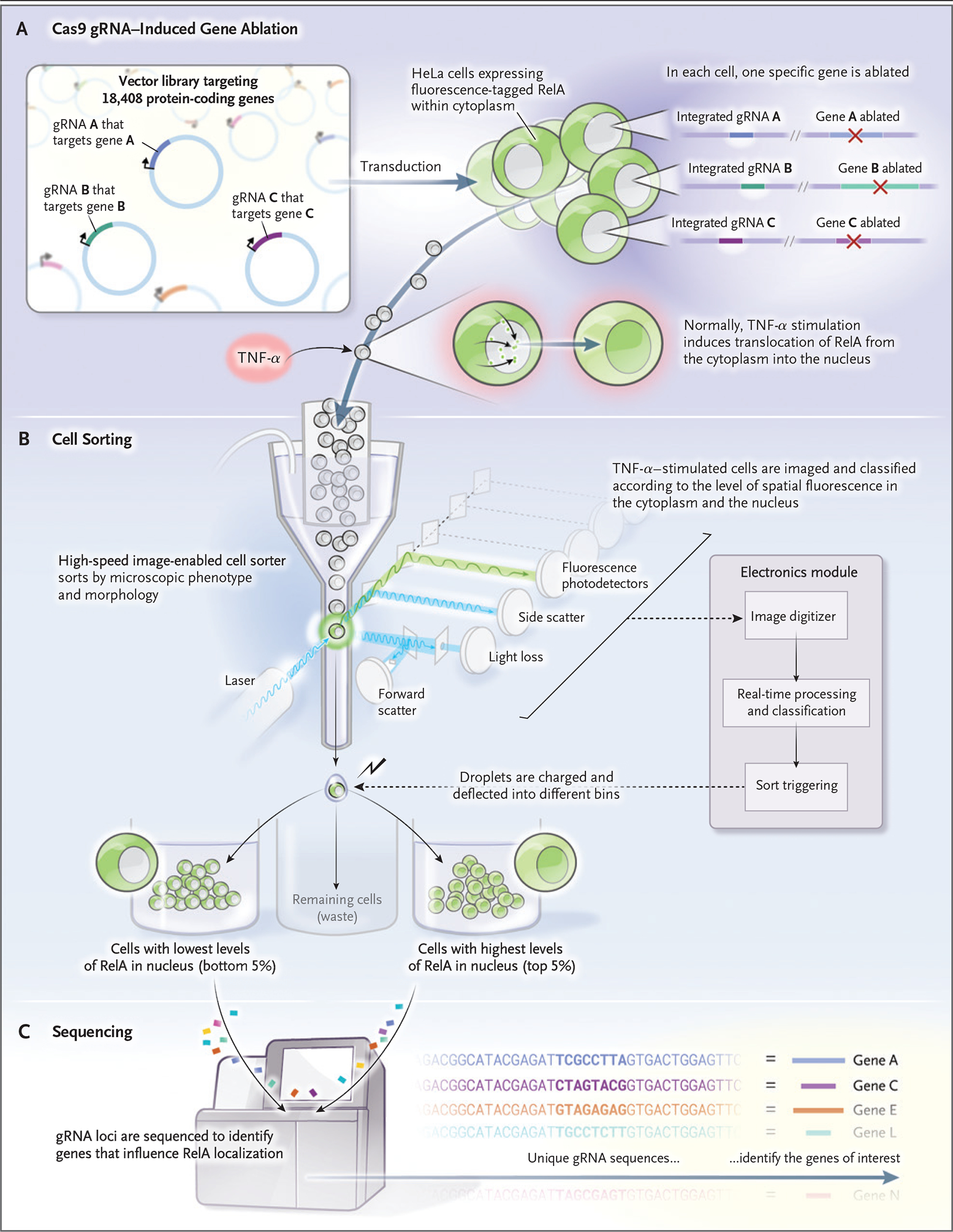
Figure 2. Genomewide Genetic Perturbation Screening.

A library of guide RNA (gRNA) is introduced into cells containing Cas9 — in this case, HeLa cells that express a fluorescent protein-tagged RelA. RelA, which normally is located in the cytoplasm, is induced to move into the nucleus when stimulated with tumor necrosis factor α (TNF-α). The stimulated cells are then sorted on the basis of correlation between the RelA intensity signal and a fluorescent nuclear stain. The 5% lowest-correlating and highest-correlating cells are physically separated from the remaining cells. The two cell populations undergo sequencing for detection of gRNA, which serves as a barcode to identify the genes that, when perturbed, affect RelA localization.
