. 2023 Jul 5;5(15):3914–3923. doi: 10.1039/d3na00124e

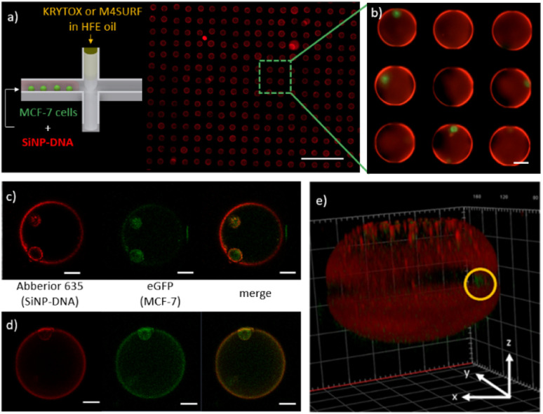
Fig. 5. (a) Schematic representation of the encapsulation of MCF-7eGFP cells with SiNP-DNA in cell culture medium (left) and a representative fluorescence image of the microfluidic chip containing an array of droplets inside the on-chip storage wells (right). The scale bar is 500 μm. (b) Enlarged image of microfluidic droplets containing MCF-7 cells and self-assembled SiNP-DNA. Scale bar is 50 μm. (c and d) 2D confocal images of the MCF-7eGFP cells (green) inside microfluidic droplets obtained with the SiNP-DNA/KRYTOX (c) and SiNP/M4SURF (d) systems. The shell-like structures (red) contain the MCF-7eGFP cells (green) that adhered to the SiNP-DNA-decorated interface. Scale bars are 25 μm. (e) 3D reconstruction image of a representative microfluidic droplet containing an encapsulated MCF-7eGFP cell (green, marked by a yellow circle) and the self-assembled SiNP-DNA layer.

Fig. 5