. 2023 Jul 4;5(15):3942–3954. doi: 10.1039/d3na00032j

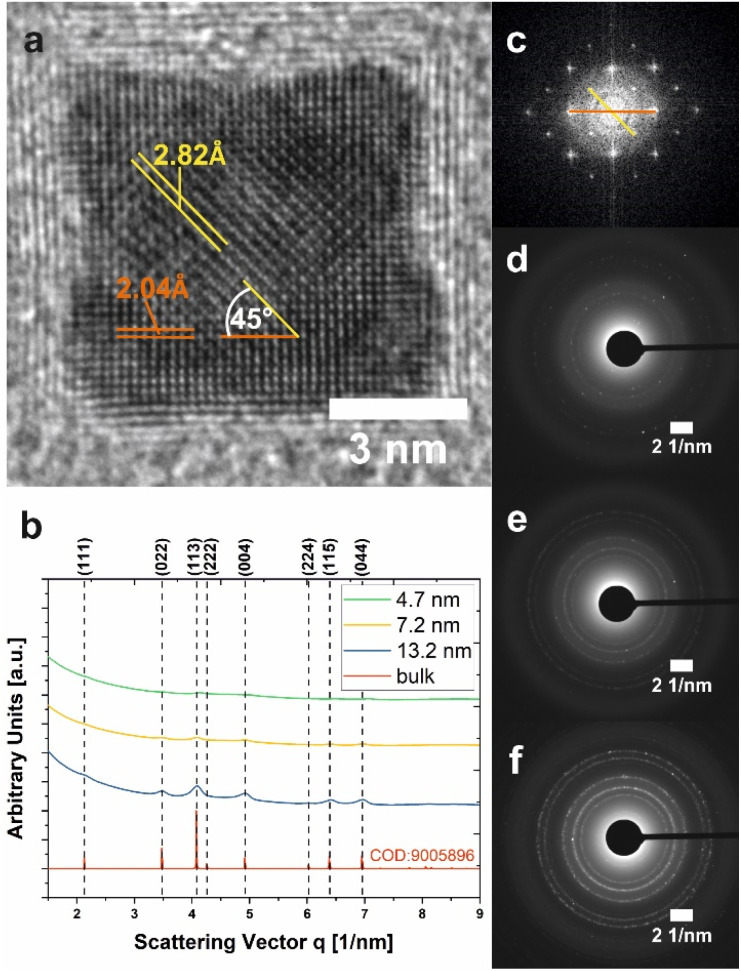
Fig. 5. Crystallographic characterization of Co3O4 NPs. (a) HR-TEM micrograph of an individual 7.2 nm-sized Co3O4 nanocube (Cb8). Two sets of lattice fringes with distances attributable to {022} and {004} lattice planes in the spinel-Co3O4 structure are highlighted in orange and yellow, respectively. (b) Electron diffractograms of samples S5 (green trace), Cb8 (yellow trace), and Cb13 (blue trace) obtained from SAED measurements. The peak profile of bulk Co3O4 is shown for comparison (red line). (c) Fast Fourier transformation corresponding to (a). (d–f) SAED patterns of NPs with 4.7 nm (S5), 7.2 nm (Cb8), and 13.2 nm (Cb13) size, respectively. The displayed data correspond to NPs in Fig. 3.

Fig. 5