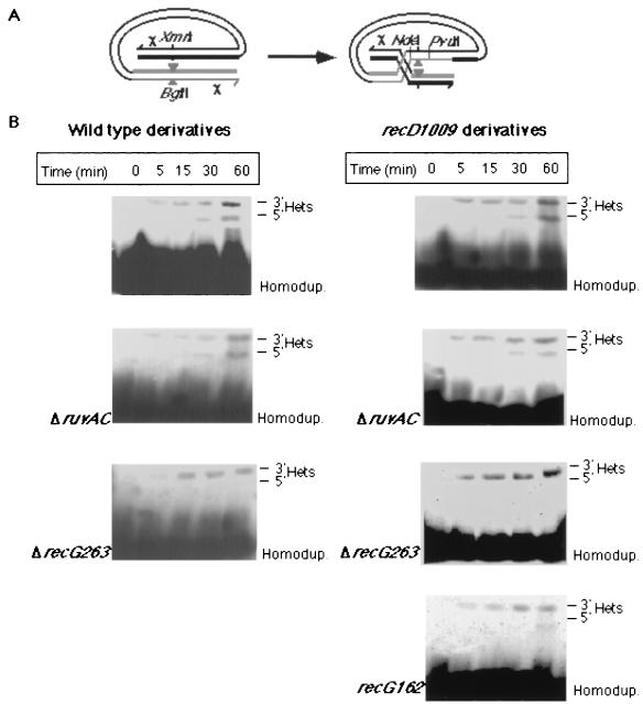
FIG. 2.
The effect of recG and ruvAC mutations on heteroduplex strand polarity. (A) A schematic representation of the phage-delivered intramolecular recombination substrate. The location of the Chi octamers (χ), relevant restriction sites, and the eight-nucleotide BglII linker (triangles) are indicated. (B) Total cellular DNA preparations (15 μg/lane) of samples taken at the indicated times after infection (MOI = 3) were subjected to Southern hybridization analysis as described in Materials and Methods. Genotypes of the infected cells are indicated. All wild-type derivatives were infected with λRF953, and all recD1009 derivatives were infected with λZS820. The locations of the electrophoretic bands of the homoduplexes (Homodup.) and the 3′ (3′ Het.) or 5′ (5′ Het.) heteroduplexes are indicated.

