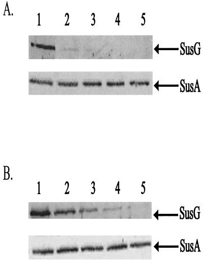
FIG. 3.
Immunoblots showing proteolytic sensitivity of SusG in intact cells. Cells were treated with proteinase K (2 mg/ml), and degradation of SusG was observed over time. SusA, a periplasmic marker, is shown to be stable during the course of each experiment. Approximately 100 μg of protein from whole-cell extracts was loaded onto each lane. Lanes 1 to 5 represent whole-cell extracts of B. thetaiotaomicron wild-type (A) and ΩsusC(pSGC23A) (B) strains at 0, 30, 60, 120, and 240 min, respectively.

